Patient Information
See Our Reviews / Leave A Review
Endodontic Faq
What Is Endodontics?
Endodontics is a branch of dentistry recognized by the American Dental Association involving treatment of the pulp (root canal) and surrounding tissues of the tooth. When you look at your tooth in the mirror, what you see is the crown. The rest of the tooth, the portion hidden beneath the gum line, is called the root. Though the outer portion of the root is a hard tissue called dentin, the inside channel or “root canal” contains a pulp of soft tissue, blood vessels and nerves. Bacteria that are introduced into the pulp as a result of tooth decay, periodontal disease, tooth fracture or other problems, can severely damage the pulp.
When that happens, an endodontic specialist can remove the diseased pulp to save the tooth and prevent further infection and inflammation. After successful endodontic treatment, the tooth continues to feel and function as it did previously.

I’m Worried About X-Rays. Should I Be?
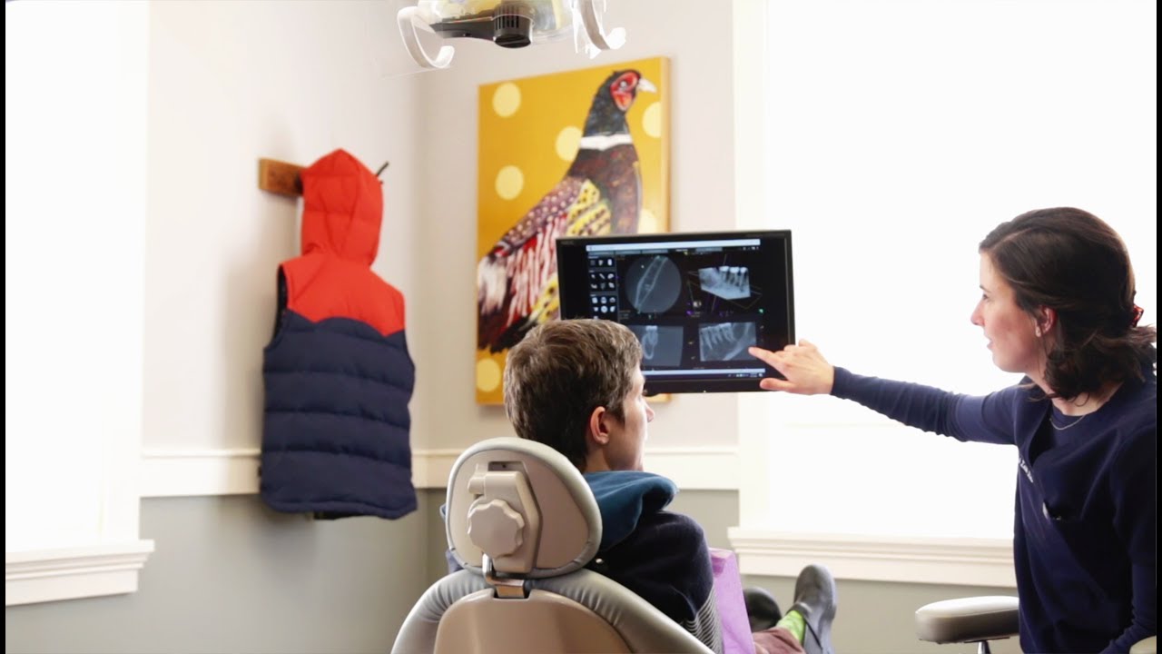
No. While x-rays will be necessary during your endodontic treatment, we use an advanced non-film computerized system, called digital radiography, that produces radiation levels up to 90 percent lower than those of already low dose conventional dental x-ray machinery. These digital images can be optimized, archived, printed, and sent to other treating doctors via e-mail.
What About Infection?
Again, there’s no need for concern. We exceed most of the rigorous standards of infection control advocated by OSHA, the Centers for Disease Control, and the American Dental Association. We utilize autoclave and steam sterilization and barrier techniques to eliminate any risk of infection.
What Happens After Root Canal Treatment?
When your root canal therapy has been completed, a record of your treatment will be sent to your restorative dentist.

You should contact his/her office for a follow-up restoration within a month of completion at our office. Your restorative dentist will decide on what type of restoration is necessary to protect your tooth. It is rare for endodontic patients to experience complications after routine endodontic treatment or microsurgery. If a problem does occur, however, we are here and available to do our best to properly resolve your problem.
What New Technologies Are Being Used?
Operating Microscopes:
In addition to digital radiography, we utilize special operating microscopes. Magnification and LED fiber optic illumination are helpful in aiding the doctor to see tiny details inside your tooth. Also, a tiny video camera on the operating microscope can record images of your tooth to further document the doctor’s findings.
Ultra-Low Radiation CBCT 3-Dimentional Imaging
Our practice utilizes a state-of-the-art, ultra-low radiation, small field cone-beam CT (computed tomography) technology by Morita that provides highly accurate, 3-D radiographic images for the diagnosis, planning, and treatment of endodontic disease. This allows for three-dimensional visualization of teeth, bone, sinuses, and surrounding structures with minimal radiation to the patient, enabling a level of anatomical accuracy and patient care not possible with 2-D technologies (regular digital dental x-rays) alone.
With the addition of cone-beam CT technology to our office, our practice is committed to providing innovative, high-quality, patient care.